Bone loss in inflammatory disorders
- R Hardy and
- M S Cooper
- School of Clinical and Experimental Medicine, Institute of Biomedical Research, University of Birmingham, Birmingham B15 2TT, UK
- (Correspondence should be addressed to M S Cooper who is now at Endocrinology, School of Clinical and Experimental Medicine, Queen Elizabeth Hospital, University of Birmingham, Edgbaston, Birmingham B15 2TH, UK; Email: m.s.cooper{at}bham.ac.uk)
-
Figure 2
The canonical Wnt pathway: the wnt pathway increases β-catenin availability. In the absence of Wnt signalling, a destruction complex containing axin, glycogen synthase kinase-3b (GSK-3b), adenomatous polypsis coli (APC) and casein kinase 1 (CK1) targets β-catenin for proteosomal degradation. Wnt signalling is initiated by binding of Wnts to the frizzled (Fz) and LRP5/6 co-receptor complex. Activation causes recruitment of GSK-3β binding protein (GBP), which interferes with β-catenin degradation. β-Catenin accumulates within the cytosol and translocates into the nuclues where it binds to TCF/LEF transcription factors and initiates transcription of Wnt target genes. Several secreted factors inhibit Wnt signalling through binding to Wnts (e.g. secreted Fz related proteins (SFrp) and Wif1) or the LRP5/6 receptor (e.g. DKK1 and sclerostin).
-
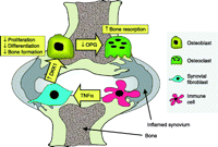
Figure 3
Schematic illustration of the possible role of DKK1 in the bone remodelling imbalance in inflammatory joint disease. Production of DKK1 in response to TNF-α production by inflammatory cells is proposed to inhibit bone formation but increase bone resorption by osteoclasts through a suppression of OPG production by osteoblasts.
- © 2009 Society for Endocrinology


