
Targeting PI3K/mTOR signaling exerts potent antitumor activity in pheochromocytoma in vivo
- Misu Lee1,*,
- Ninelia Minaskan1,
- Tobias Wiedemann1,
- Martin Irmler2,
- Johannes Beckers2,3,4,
- Behrooz H Yousefi5,
- Georgios Kaissis6,
- Rickmer Braren6,
- Iina Laitinen7,† and
- Natalia S Pellegata1⇑
- 1Institute for Diabetes and Cancer, Helmholtz Zentrum München, Neuherberg, Germany
- 2Institute of Experimental Genetics, Helmholtz Zentrum München, Neuherberg, Germany
- 3German Center for Diabetes Research (DZD), Neuherberg, Germany
- 4Technische Universität München, Chair of Experimental Genetics, Freising, Germany
- 5Department of Pharmaceutical Radiochemistry, Technische Universität München, Garching, Germany
- 6Institute for Diagnostic and Interventional Radiology, Klinikum rechts der Isar der Technische Universität München, Munich, Germany
- 7Department of Nuclear Medicine, Klinikum rechts der Isar der Technische Universität München, Munich, Germany
- Correspondence should be addressed to N S Pellegata; Email: Natalia.pellegata{at}helmholtz-muenchen.de
-
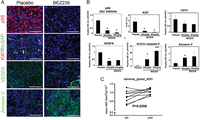 Figure 1
Analysis of PCC tissues from rats treated with placebo or BEZ235. (A) Tumor tissues of MENX-affected rats treated with PEG vehicle (Placebo) or with 30 mg/kg of BEZ235 by oral gavage for 2 weeks. Tissues were collected, fixed in formalin and embedded in paraffin. Immunofluorescent (IF) staining was performed using specific antibodies raised against P-S6 (Ser240/244), Ki67, NuSAP, VEGFA and Annexin V. Cell nuclei were counterstained with DAPI. Scale bars: 50 µm. (B) Quantification of the IF signal of the panels shown in A and in Supplementary Fig. 2 done with ImageJ software for the antibodies against P-S6, CD31, VEGFA, active caspase-3 and Annexin V. For Ki67, the percentage of the positive nuclei was counted in 5× high-power field (HPF) on tissues from 3 different mutant rats for each condition. *P < 0.05, **P < 0.01 (C) Apparent diffusion coefficient (ADC) values were obtained by performing DW-MRI before (pre) and after (post) treatment of mutant rats (n = 5, total adrenal gland number n = 10) with BEZ235.
-
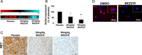 Figure 2
BEZ235 reduces Slc6a2/NET expression in rat PCCs in vivo and in vitro. (A) Heat map showing the relative expression of Slc6a2 obtained by gene expression profiling of PCCs of rats treated with placebo (n = 8) or BEZ235 (20 mg/kg, n = 5; 30 mg/kg, n = 4) for 14 days. (B) RNA was extracted from the PCCs of the rats used in A. qRT-PCR was performed using TaqMan primer and probe sets specific to rat Slc6a2. The relative mRNA expression level of Slc6a2 was calculated with the 2−ΔΔCt formula and was normalized for input RNA using rat β2-microglobulin gene expression (housekeeping gene). The obtained relative value was normalized against the average expression of placebo-treated tissues arbitrarily set to 100. Data were analyzed independently with six replicates each and are expressed as the mean ± s.e.m. *P < 0.05. (C) Rat PCCs were collected after 14 days of daily placebo or BEZ235 administration and was analyzed by immunostaining for NET expression. Scale bars: 20 µm. (D) In vitro cultures of rat primary PCC cells were treated daily with DMSO vehicle or with BEZ235 (1 µM) for 48 h. Then, they were fixed and immunofluorescent staining was conducted using the anti-NET antibody. Nuclei were counterstained with DAPI. Scale bars: 50 µm.
-
Figure 3
The expression of Slc6a2/NET in PCC cells. (A) Semi-quantitative RT-PCR and western blotting specific to the Slc6a2 gene/NET protein, respectively, were performed using 3 PCC cell lines (PC12, MPC and MTT). As internal control for the mRNA level we used Gadph, whereas for the protein level, we used α-tubulin. (B) MPC cells were incubated with DMSO (vehicle) or with 1 µM BEZ235. RNA and proteins were extracted 48 h later. RT-PCR was performed to monitor Slc6a2 expression as in A. In parallel, NET, AKT, P-AKT (Ser473), S6 and P-S6 (Ser240/244) expression was monitored by western blotting. (C) MPC cells were incubated with 1 µM BEZ235, 1 µM BKM120, 1 µM RAD001 or with the combination BKM120 and RAD001. Control cells were incubated with DMSO (vehicle). RNA was extracted 48 h later, and TaqMan analysis for the Slc6a2 gene was performed. Shown is the average of the relative expression values were normalized against the β2-microglobulin housekeeping gene and was calculated with the 2−ΔΔCt formula. The obtained relative value was normalized against the average expression of DMSO-treated cells arbitrarily set to 1. Data were analyzed independently with three replicates each and are expressed as the mean ± s.e.m. *P < 0.05. (D) From samples parallel to C, proteins were extracted after drug treatment, and the expression of NET, P-AKT (Ser473) and P-S6 (Ser240/244) was monitored by western blotting.
-
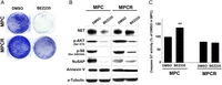 Figure 4
The expression of NET in MPC and MPCR cells after BEZ235 treatment. (A) Colony formation assay of MPC and MPCR cells treated with DMSO (vehicle) or with 1 µM BEZ235 for 2 weeks. Cells were then fixed and stained with crystal violet. Representative plates are shown. (B) MPC and MPCR cells were incubated with DMSO (vehicle) or with 1 µM BEZ235 for 48 h. Proteins were extracted and the expression of NET, p-AKT (Ser473), p-S6 (Ser240/244), NuSAP, Annexin 5 and α-tubulin was detected by western blotting. (C) MPC and MPCR cells were treated as in B and then were monitored for caspase-3/7 activity. Shown is the average of the relative activity normalized against the DMSO-treated cells arbitrarily set to 100. Data were analyzed independently with six replicates each and were expressed as the mean ± s.e.m. **P < 0.01.
-
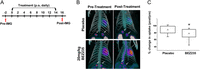 Figure 5
Functional imaging with 18F-LMI1195 to monitor the response of rat PCCs to BEZ235 in vivo. (A) Scheme of the treatment course. PET imaging (IMG) was performed before (pre) and after (post) treatment with placebo (PEG) or BEZ235 for 14 days. (B) Representative images of the PET scans obtained for the same mutant rats before and after treatment with placebo or BEZ235. Arrows indicate the adrenal glands. (C) Quantitative analysis of PET images of 18F-LMI1195 in placebo-treated (n = 5; 10 adrenals) and BEZ235-treated MENX rats (n = 8; 16 adrenals).
- © 2017 Society for Endocrinology


