
Hyperprolactinemia induced by hCG leads to metabolic disturbances in female mice
- Laura D Ratner1,
- Guillermina Stevens1,2,
- Maria Marta Bonaventura1,
- Victoria A Lux-Lantos1,
- Matti Poutanen3,4,
- Ricardo S Calandra1,
- Ilpo T Huhtaniemi3,5 and
- Susana B Rulli1⇑
- 1Instituto de Biología y Medicina Experimental- Consejo Nacional de Investigaciones Científicas y Técnicas, Buenos Aires, Argentina
- 2Hospital General de Agudos J. M. Ramos Mejía, Buenos Aires, Argentina
- 3Department of Physiology, Institute of Biomedicine, University of Turku, Turku, Finland
- 4Turku Center for Disease Modeling, University of Turku, Turku, Finland
- 5Department of Surgery and Cancer, Imperial College London, London, UK
- Correspondence should be addressed to S B Rulli; Email: srulli{at}ibyme.conicet.gov.ar
-
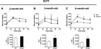 Figure 1
Intraperitoneal glucose tolerance test (IGTT) in hCGβ+ female mice at 2, 3, and 6 month of age. IGTT (2g/kg) in fasted WT and hCGβ+ females was performed at 2 (A), 3 (B) (n=4), and 6 (C) months of age (n=7). Two-way ANOVA with repeated measures, followed by Bonferroni’s post hoc test, was conducted; *P<0.05; **P<0.01; ***P<0.001. The area under the curve (AUC) was analyzed for each group at different ages; Student’s t-test was conducted; **P<0.01. Data are presented as mean±s.e.m.
-
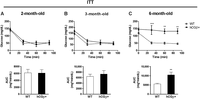 Figure 2
Intraperitoneal insulin tolerance test (ITT) in hCGβ+ female mice at 2, 3, and 6 month of age. ITT (0.75IU/kg) in fasted WT and hCGβ+ females was performed at 2 (A), 3 (B) (n=4), and 6 (C) months of age (n=6). Two-way ANOVA with repeated measures, followed by Bonferroni’s post hoc test, was conducted; **P<0.01; ***P<0.001. The area under the curve (AUC) was analyzed for each group at different ages; Student’s t-test was conducted; **P<0.01. Data are presented as mean±s.e.m.
-
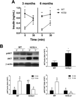 Figure 3
(A) Glucose-stimulated insulin release in 3- and 6-month-old hCGβ+ female mice. Glucose (2g/kg) was administered i.p. to fasted WT and hCGβ+ female mice, and serum insulin levels were measured at 0 and 30min after glucose administration. Two-way ANOVA with repeated measures, followed by Bonferroni’s post hoc test, was conducted. Data are presented as mean±s.e.m. (n=4). *P<0.05; **P<0.01. (B) Peripheral tissue response to insulin. A representative western blot was shown for AKT activation in skeletal muscle. Samples were obtained from fasted 6-month-old WT and hCGβ+ female mice at 0 or 5 min after insulin administration. Two-way ANOVA, followed by Bonferroni’s post hoc test or Student’s t-test, was conducted according to each case. Different letters indicate a value of at least P<0.05. Data are presented as mean±s.e.m. (n=4).
-
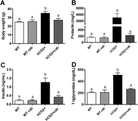 Figure 4
Effect of cabergoline on body weight and serum levels of prolactin, insulin, and triglycerides in hCGβ+ females. Body weight (A) and serum prolactin (B), insulin (C) and trygicerides (D) levels in 6-month-old cabergoline-treated transgenic females (hCGβ+cab) (n=8) after 18h fasting. WT (n=12), cabergoline-treated WT (WTcab) (n=4), and hCGβ+ (n=8) females were used as controls. ANOVA, followed by Bonferroni’s post hoc test, was conducted. Different letters indicate a value of at least P<0.05. Data are presented as mean±s.e.m.
-
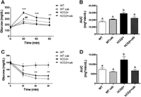 Figure 5
Effect of cabergoline treatment on the glucose homeostasis in WT and hCGβ+ females. IGTT (2g/kg) (A) and ITT (0.75IU/kg) (C) in fasted 6-month-old cabergoline-treated transgenic (hCGβ+cab) females was performed (n=8); fasted 6-month-old WT (n=12), cabergoline-treated WT (WTcab) (n=4), and hCGβ+ females (n=12) were used as control groups. Two-way ANOVA with repeated measures, followed by Bonferroni’s post hoc test, was conducted. (A) WT vs hCGβ+ **P<0.01; ***P<0.001; hCGβ+ vs hCGβ+cab ##P<0.01; (C) hCGβ+ vs WT, WTcab, hCGβ+cab; **P<0.01, ***P<0.001. The area under the curve (AUC) was analyzed for the different groups (B and D). ANOVA, followed by Bonferroni’s post hoc test, was conducted. Different letters indicate a value of at least P<0.05. Data are presented as mean±s.e.m.
-
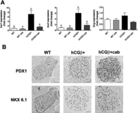 Figure 6
(A) Effect of cabergoline on the pancreatic gene expression of Ins1, Ins2, Gcg, and Ccnd2. The mRNA expression analysis of Ins1, Ins2, and Gcg from fasted 6-month-old WT, hCGβ+, and cabergoline-treated WT (WTcab), and hCGβ+ (hCGβ+cab) pancreata was carried out by qPCR. ANOVA, followed by Bonferroni’s post hoc test, was conducted. Different letters indicate a value of at least P<0.05. Data are presented as mean±s.e.m. (n=4). (B) immunolocalization of PDX1 and NKX 6.1 in pancreas. Representative sections from 6-month-old WT, hCGβ+, and hCGβ+cab female mice were immunostained with PDX1- and NKX 6.1-specific antibodies. Scale bar 100 µm.
- © 2016 Society for Endocrinology


