
Enhanced intestinal epithelial cell proliferation in diabetic rats correlates with β-catenin accumulation
- Tatiana Dorfman1,
- Yulia Pollak1,
- Rima Sohotnik1,
- Arnold G Coran4,
- Jacob Bejar3 and
- Igor Sukhotnik1,2⇑
- 1Laboratory of Intestinal Adaptation and Recovery, The Ruth and Bruce Rappaport Faculty of Medicine, Technion‐Israel Institute of Technology, Haifa, Israel
2Departments of Pediatric Surgery B
3Pathology, Bnai Zion Medical Center, 47 Golomb Street, PO Box 4940, Haifa 31048, Israel
4Section of Pediatric Surgery, C.S. Mott Children's Hospital, University of Michigan Medical School, Ann Arbor, Michigan, USA
- Correspondence should be addressed to I Sukhotnik; Email: igor-dr{at}internet-zahav.net
-
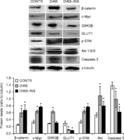 Figure 1
Relative expression of β-catenin, caspase 3, GSK3β, c-Myc, ERK, Akt, and GLUT1 protein, as determined by western blot analysis. A significant upregulation of Wnt/β-catenin-signaling-related genes in diabetic animals was accompanied by a significant increase in β-, c-Myc (target), GSK3β, Akt, and p-ERK protein levels compared with those for control animals. Decreased rates of cell apoptosis in diabetic rats correlated with a significant decrease in caspase 3 protein levels compared with those of control animals. Insulin-treated rats demonstrated a trend towards a decrease in GSK3β and Akt levels; however, this trend was not statistically significant. CONTR, control rats; DIAB, diabetic rats; INS, insulin. Values are mean±s.e.m. *P<0.05 DIAB and DIAB–INS versus CONTR.
-
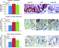 Figure 2
Effect of diabetes and insulin on intestinal expression of β-catenin, Musashi 1, and c-Myc (immunohistochemistry). As expected, diabetic rats demonstrated a significant increase in the intensity of Musashi 1 immunostaining compared with that for control animals, indicating elevated stem cell activity. In addition, diabetic rats demonstrated higher β-catenin staining in cytoplasm and nucleus as well as an increased number of c-Myc-positive cells (nuclear staining (arrows)) compared with the results for control rats. Treatment with insulin resulted in a significant decrease in the intensity of Musashi 1 immunostaining compared with that for sham-treated animals, indicating decreased stem cell activity, which was accompanied by a trend towards a decrease in the number of β-catenin- and c-Myc-positive cells (nuclear staining (arrows)); however, this trend was not statistically significant. CONTR, control rats; DIAB, diabetic rats; INS, insulin. Values are mean±s.e.m. *P<0.05 DIAB and DIAB–INS versus CONTR and †P<0.05 DIAB–INS versus DIAB.
- © 2015 Society for Endocrinology


