The skeleton: a multi-functional complex organ. The growth plate chondrocyte and endochondral ossification
- (Correspondence should be addressed to E J Mackie; Email: ejmackie{at}unimelb.edu.au)
-
Figure 1
Overview of development of a bone by endochondral ossification. (A) The cartilage model of the future bone. (B) A periosteal bone collar has formed and formation of the primary centre of ossification has been initiated. (C) The primary centre of ossification is starting to expand towards the ends of the cartilage model. (D) The secondary centres of ossification have formed at each end of the bone, leaving a cartilaginous growth plate between primary and secondary centres of ossification. (E) Skeletal maturity has been achieved, with complete replacement of the growth plate cartilage by bone. The only cartilage remaining is the articular cartilage at the ends of the bone.
-
Figure 3
The changing morphology of growth plate chondrocytes. Chondrocytes in a section through the proximal growth plate of a 4-week-old rat tibia, examined by transmission electron microscopy. (A) Zone of proliferative chondrocytes, where pairs of cells that have recently undergone proliferation have not yet separated from each other. (B) Zone of early hypertrophic chondrocytes, where ECM now separates the cells. (C) Zone of late hypertrophic chondrocytes, including the last lacunae before the ossification front, which can be identified by the presence of erythrocytes (arrow). The majority of cells are light (li) chondrocytes, but occasional dark (da) chondrocytes are visible in all zones. All figure parts are the same magnification; bar=10 μm.
-
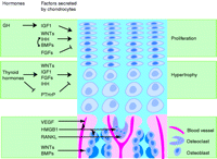 Figure 4
Overview of the effects of factors secreted by chondrocytes on growth plate cell function and invasion. Under the control of circulating hormones, chondrocytes secrete growth factors that act on chondrocytes to regulate their proliferation and hypertrophy (upper two panels), and on cells of the ossification front (lower panel), to regulate their invasion of the growth plate cartilage. Arrows indicate stimulatory pathways, and crossed lines indicate inhibitory pathways. Not all effects mentioned in the text are included in this figure.
- © 2011 Society for Endocrinology