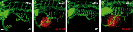
MTC-grafted cells stimulate angiogenesis in zebrafish embryos. Representative confocal microscopic images of Tg(fli1:EGFP)y1 zebrafish embryos implanted with red fluorescence-stained MZ-CRC1 (B and D) cells. 24 (A and B) and 48 (C and D) hours after injection (hpi), larvae were embedded in low-melting agarose, and the yolk region was observed by confocal microscopy. In comparison to control larvae (A and B), MTC-grafted larvae showed endothelial structures (green) that sprout from the subintestinal vein (SIV) plexus and the common cardinal vein (CCV) (B and D) and progressively form new vessels. All images are oriented so that rostral is to the left and dorsal is at the top. Scale bar, 50 μm.